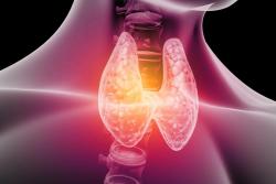
The gallbladder often doesn’t get the recognition it deserves as it can be seen as just a storage component for bile, and the body is able to function quite well without it. While it is true that digestive functions can still work without a gallbladder, diseases of the gallbladder are often multifactorial, and both bile flow and gallbladder function play a big part in overall health. Often, individuals may struggle with fat digestion and suffer with symptoms such as bloating, abdominal pain and reflux, which leads them to follow a low-fat, or generally restricted diet. However, there are a lot of diet and lifestyle changes that can be made to help reduce symptoms and improve quality of life in people with gallbladder issues.
Why is the Gallbladder Important?
The main role of the gallbladder is to store and concentrate bile that is produced by the liver, and due to this, it’s located just under the liver with the two organs are connected by the biliary tract. Together, they help to facilitate digestion and aid the absorption of fats from the food that we eat.
The liver uses cholesterol to create the primary bile acids; cholic acid & chenodeoxycholic acid. The liver can then attach amino acids glycine and taurine to these bile acids to create 8 different bile salts, which make up the main component of bile, alongside cholesterol, phospholipids, water, proteins and conjugated bilirubin.1 Bile is pushed through the common hepatic duct to the gallbladder where it is concentrated and stored. In the presence of fat within a meal, cholecystokinin (CCK) is released to stimulate the contraction of the gallbladder. This squeezes bile out of the gallbladder, through the cystic duct and into the duodenum where it helps to emulsify fats into micelles, which are smaller molecules that pancreatic lipase can work on. Essential fats and fat-soluble nutrients such as vitamins A, D, E and K found in the food we eat are absorbed through this process.2
In addition to these main functions, bile also plays an important role in the removal of toxins and waste products from the body, such as excess bilirubin and cholesterol, inhibiting growth of harmful microorganisms, and stimulating colonic motility.3
Common Gallbladder Issues
There are a few conditions that affect the function of the gallbladder, with the most common being the formation of gallstones.4 Gallstones are made from components of bile, creating two main types of gallstones which both have their own unique pathophysiology and risk factors. The most common form of stones are cholesterol stones, which can develop when bile contains too much cholesterol, bilirubin, or insufficient amounts of bile salts or choline. 5 Also, gallbladder stasis, which is characterized by reduced bile flow and stagnation, can increase the risk of cholesterol gallstone formation. Brown, or pigmented stones are less common, but are caused by bacteria such as E.coli which can release enzymes that hydrolyse conjugated bilirubin and phospholipids, which can then bind with calcium ions and cause stone formation. 6
- Factors that can increase the risk of developing gallstones include:
- Family history of gallstones or gallbladder disease
- Obesity
- Gender (more common in women)
- Increased levels of oestrogen
- Pregnancy
- Being over forty years of age. 7
Women are at a higher risk of gallstones as higher levels of oestrogen can increase cholesterol saturation within bile, which contributes to cholesterol stones.8 Increased levels of progesterone can also impair gallbladder emptying in response to CCK, contributing to bile stasis.9 Because of these mechanisms, women on hormonal therapies and contraception may have a higher risk of gallstone formation.
Biliary colic (also known as a gallbladder attack), happens when a gallstone in the gallbladder gets lodged in the bile duct, due to the gallbladder contracting in response to a meal containing fat and moving the stone. This causes temporary, severe abdominal pain, which can last around 6 hours, until the gallstone dislodges itself and falls back into the gallbladder.5
Symptoms of gallstones and other gallbladder dysfunction include:
- Pain under the ribs which can radiate to the right shoulder or back
- Nausea
- Abdominal pain
- Vomiting
- Fever
- Indigestion
- Pale-coloured stools
- Floating/fatty stools
- Diarrhoea. 10
What can affect fat digestion & bile flow?
Liver dysfunction
As the liver is the organ that creates bile, any damage that occurs to hepatocytes can result in reduced bile production or cholestasis, whereby the flow of bile out of the liver is disrupted.4 This can lead to an accumulation of bile acids within the liver and increased pro-inflammatory cytokines, which can lead to death of the hepatocytes. 11 Cholestasis can be caused by genetic, diet, lifestyle & medication factors such as drug-induced liver toxicity, alcohol, viral hepatitis, autoimmune destruction of bile ducts, factors associated with pregnancy, or obstruction of the bile duct due to something like a gallstone or tumor. 12
Farnesoid X Receptor’s influence on metabolism
The Farnesoid X Receptor (FXR) plays an important role in regulating multiple metabolic pathways including glucose, lipid and energy homeostasis, as well regulating the synthesis, transport and reabsorption of bile acids. 22 These receptors are mostly present within the liver and ileum and are activated by bile acids. Disruption of the bile acid pool, or the gut microbiome can interfere with optimal FXR signaling, which can play a role in the development of NAFLD, 23 gut dysbiosis, metabolic dysfunction and inflammation. 24–26
The role of the gut microbiome
Our gut microbiome can interact with primary bile acids as they move through the intestines, and this produces secondary bile acids. Normally, 95% of bile acids should be reabsorbed via enterohepatic circulation where they are sent back to the liver to be recycled, 13 leaving a very small percentage of around 5% of bile acids being lost in feces. 14 However, if there is damage to the small intestine through factors such as autoimmunity, dysbiosis, coeliac disease, IBD and cancer treatments, the passive absorption of bile acids can be affected, decreasing the amount that are reabsorbed, which can contribute to a bile acid deficiency. 15 In some cases, it can cause a buildup of bile acids within the intestines, irritating the colon and resulting in chronic diarrhea and loose stools.
In addition, if bile acids are not being reabsorbed and recycled optimally, it can leave more bile acids available for the gut bacteria to interact with. 16 An overgrowth of bacteria within the small intestine can be detrimental as some of the secondary bile acids have been linked to inflammation and chronic diseases such as colon cancer. 13,16 In case of small intestinal bacterial overgrowth (SIBO), the bacteria within the small intestine can cause damage to the small intestine and deconjugate bile acids, leading to bile acid malabsorption. 17,18 On the contrary, a healthy microbiome, can produce beneficial secondary bile acids that can assist in regulating body weight and lipid metabolism,19 absorption of nutrients, modulating the immune system 20 and aiding the integrity of the gut. 21
The impact of bile on the gut microbiome
There is a bidirectional link between bile and gut bacteria. Bile acids can offer protection against bacterial proliferation in the small intestine through the activation of FXR and gene expression, which also help to support the epithelial integrity. 27 Activation of FXR by conjugated bile acids induces the expression of genes responsible for preventing bacterial overgrowth and promoting epithelial integrity. 28 Bile itself has antimicrobial properties thanks to its ability to disrupt cell membranes of both gram-positive and gram-negative bacteria ,29,30 which makes it vital for regulating a healthy composition of the microbiome and preventing dysbiosis, including SIBO. 31,32,33
What you can do
Support bile production & flow
Liver function is essential for bile production, so offering support to the liver is essential. Artichoke, 34 milk thistle 35 and turmeric 36 are hepaprotective and promote bile flow for fat digestion and cholesterol clearance.
Phosphatidylcholine is an important nutrient to support liver function, 37,38 fat transport and metabolism, gut integrity, bile flow, and may help to dissolve gallstones. 39 The PEMT gene has the job of producing phosphatidylcholine using SAMe as a methyl donor group. However, variants of the PEMT gene can contribute to a choline deficiency and increase the risk of NAFLD, gallbladder issues, and raised triglycerides. 40,41 For PEMT to function optimally, it requires sufficient amounts of SAMe, produced through the methionine cycle, alongside choline, therefore it’s important to increase dietary and supplemental sources of methylfolate and methylcobalamin in particular to promote methylation.
Foods high in choline include liver, eggs, fish & meat, which can make it more difficult to reach choline needs on a vegan or vegetarian diet. 42 There are vegetables that can be beneficial include broccoli, cauliflower, spinach, lentils, beetroot and shiitake mushrooms, but choline supplementation may be needed. 43
Promote a healthy gut microbiome
If an individual is struggling with gallbladder issues, or issues with fat digestion, repairing the gut and supporting the gut microbiome needs to be considered. Increasing fibre and prebiotics can help to act as a bulking agent in the bowel to encourage peristaltic motion helping with both diarrhoea and constipation. 44,45 As well, both prebiotics and probiotics can promote beneficial bacteria in the colon and reduce growth of potentially pathogenic bacteria including Escherichia coli. 46 In addition, both prebiotics and probiotic can help improve metabolic function through regulating glucose metabolism, 47,48 and cholesterol levels, 49 which may reduce risk factors for gallstones. 50
Support post gallbladder removal
Chronic gallbladder issues can result in a cholecystectomy (removal of the gallbladder). This is an essential surgery if bile ducts have become blocked, which can cause the gallbladder to become inflamed. However, once the gallbladder is removed there is no longer storage space for bile, which means that bile continuously drips into the duodenum, instead of larger amounts being secreted with meals. Digestion and absorption of foods will still function well after a cholecystectomy, but some individuals may find they struggle with meals higher in fats or fibre. In the long-term, a cholecystectomy can contribute to certain nutrient deficiencies, especially fat-soluble vitamins 51 due to more diluted and lower concentrations of bile, as well as diarrhoea, fatty liver, and reduced microbiome diversity. 52,53
Unfortunately, even when the gallbladder has been removed, there can still be a risk of gallstones formation within the biliary tree or liver. 54 In the presence of gallstones, or gallbladder removal, digestive enzymes such as lipase may help to reduce symptoms related to poor digestion of fats. 55 In those cases, small amounts of herbs that promote liver health and bile production may be recommended before meals, ideally alongside a high dose of lipase enzymes, but high doses should be taken cautiously to prevent overstimulation of the liver.
Reduce the risk of gallstone development
One mineral that may play a significant role in reducing the risk of gallstones is magnesium. Magnesium has been found to regulate glucose metabolism and reduce raised cholesterol & triglyceride levels, all of which are risk factors for stone formation. 56 As well, research has found that diets higher in magnesium correlate which a reduced risk of gallstone development. 57 Forms such as magnesium taurate may offer an additional benefit as taurine is needed to create bile acids and healthy bile flow. 58
A Western-style diet, which is high in refined carbohydrates and sugars has been linked with an increased risk of gallstone formation. 59 Reducing the intake of processed foods, simple carbohydrates and sugars, while increasing plant foods and good quality fats and proteins may help to reduce this risk.
By identifying and working on some of the underlying factors of gallbladder dysfunction, diet and lifestyle changes can help to support all of our digestive organs and reduce the risk of disease. But, if you would like to chat through a client case study, or get some more personalised advice for yourself, please feel free to contact our Nutrition Team via clinicalnutrition@biocare.co.uk, or call +44 (0) 121 433 3727.
References
1. Hundt M, Basit H, John S. Physiology, Bile Secretion. StatPearls. Published online September 26, 2022. Accessed October 31, 2022. https://www.ncbi.nlm.nih.gov/books/NBK470209/
2. Almajid AN, Sugumar K. Physiology, Bile. StatPearls. Published online September 12, 2022. Accessed October 31, 2022. https://www.ncbi.nlm.nih.gov/books/NBK542254/
3. Ticho AL, Malhotra P, Dudeja PK, Gill RK, Alrefai WA. Bile acid receptors and gastrointestinal functions. Liver Res. 2019;3(1):31-39. doi:10.1016/j.livres.2019.01.001
4. Acalovschi M. Gallstones in patients with liver cirrhosis: Incidence, etiology, clinical and therapeutical aspects. World Journal of Gastroenterology : WJG. 2014;20(23):7277. doi:10.3748/WJG.V20.I23.7277
5. Tanaja J, Lopez RA, Meer JM. Cholelithiasis. Indian Journal of Practical Pediatrics. 2022;20(2):101-106. doi:10.5005/jp/books/12296_29
6. Soloway RD, Trotman BW, Ostrow JD. Pigment Gallstones. Gastroenterology. 1977;72(1):167-182. doi:10.1016/S0016-5085(77)80323-5
7. Bass G, Gilani SNS, Walsh TN. Validating the 5Fs mnemonic for cholelithiasis: time to include family history. Postgrad Med J. 2013;89(1057):638-641. doi:10.1136/POSTGRADMEDJ-2012-131341
8. Cirillo DJ, Wallace RB, Rodabough RJ, et al. Effect of Estrogen Therapy on Gallbladder Disease. JAMA. 2005;293(3):330-339. doi:10.1001/JAMA.293.3.330
9. Tierney S, Nakeeb A, Wong O, et al. Progesterone alters biliary flow dynamics. Ann Surg. 1999;229(2):205. doi:10.1097/00000658-199902000-00007
10. Gutt C, Schläfer S, Lammert F. The Treatment of Gallstone Disease. Dtsch Arztebl Int. 2020;117(9):148. doi:10.3238/ARZTEBL.2020.0148
11. Santiago P, Scheinberg AR, Levy C. Cholestatic liver diseases: new targets, new therapies. Therap Adv Gastroenterol. 2018;11. doi:10.1177/1756284818787400
12. Mazokopakis EE, Papadakis JA, Kofteridis DP. Unusual causes of intrahepatic cholestatic liver disease. World Journal of Gastroenterology : WJG. 2007;13(12):1879. doi:10.3748/WJG.V13.I12.1879
13. Chiang JYL. Bile Acid Metabolism and Signaling. Compr Physiol. 2013;3(3):1191. doi:10.1002/CPHY.C120023
14. Ramírez-Pérez O, Cruz-Ramón V, Chinchilla-López P, Méndez-Sánchez N. The role of the gut microbiota in bile acid metabolism. Ann Hepatol. 2017;16:S21-s26. doi:10.5604/01.3001.0010.5494
15. Camilleri M. Bile Acid Diarrhea: Prevalence, Pathogenesis, and Therapy. Gut Liver. 2015;9(3):332. doi:10.5009/GNL14397
16. Yang R, Qian L. Research on Gut Microbiota-Derived Secondary Bile Acids in Cancer Progression. Integr Cancer Ther. 2022;21. doi:10.1177/15347354221114100/ASSET/IMAGES/LARGE/10.1177_15347354221114100-FIG2.JPEG
17. Dukowicz AC, Lacy BE, Levine GM. Small Intestinal Bacterial Overgrowth: A Comprehensive Review. Gastroenterol Hepatol (N Y). 2007;3(2):112. Accessed October 31, 2022. /pmc/articles/PMC3099351/
18. Shindo K, Machida M, Koide K, Fukumura M, Yamazaki R. Deconjugation ability of bacteria isolated from the jejunal fluid of patients with progressive systemic sclerosis and its gastric pH. Hepatogastroenterology. 1998;45(23):1643-1650. Accessed October 31, 2022. https://europepmc.org/article/med/9840121
19. Ramírez-Pérez O, Cruz-Ramón V, Chinchilla-López P, Méndez-Sánchez N. The Role of Bile Acids in Glucose Metabolism and Their Relation with Diabetes. Ann Hepatol. 2017;16:S15-S20. doi:10.5604/01.3001.0010.5494
20. Yoshikawa M, Tsujii T, Matsumura K, et al. Immunomodulatory effects of ursodeoxycholic acid on immune responses. Hepatology. 1992;16(2):358-364. doi:10.1002/HEP.1840160213
21. Sun R, Xu C, Feng B, Gao X, Liu Z. Critical roles of bile acids in regulating intestinal mucosal immune responses. Therap Adv Gastroenterol. 2021;14. doi:10.1177/17562848211018098
22. Navab M, Anantharamaiah GM, Reddy ST, et al. The Farnesoid X Receptor. Arterioscler Thromb Vasc Biol. 2005;25(10):2020-2031. doi:10.1161/01.ATV.0000178994.21828.A7
23. Chow MD, Lee YH, Guo GL. The Role of Bile Acids in Nonalcoholic Fatty Liver Disease and Nonalcoholic Steatohepatitis. Mol Aspects Med. 2017;56:34. doi:10.1016/J.MAM.2017.04.004
24. Li T, Chiang JYL. Bile Acid Signaling in Metabolic Disease and Drug Therapy. Pharmacol Rev. 2014;66(4):948. doi:10.1124/PR.113.008201
25. Staels B, Fonseca VA. Bile Acids and Metabolic Regulation: Mechanisms and clinical responses to bile acid sequestration. Diabetes Care. 2009;32(Suppl 2):S237. doi:10.2337/DC09-S355
26. Chiang JYL. Bile acid metabolism and signaling in liver disease and therapy. Liver Res. 2017;1(1):3-9. doi:10.1016/j.livres.2017.05.001
27. Hofmann AF, Eckmann L. How bile acids confer gut mucosal protection against bacteria. Proc Natl Acad Sci U S A. 2006;103(12):4333. doi:10.1073/PNAS.0600780103
28. Mori H, Svegliati Baroni G, Marzioni M, et al. Farnesoid X Receptor, Bile Acid Metabolism, and Gut Microbiota. Metabolites. 2022;12(7). doi:10.3390/METABO12070647
29. Tam J, Icho S, Utama E, et al. Intestinal bile acids directly modulate the structure and function of C. Difficile TcdB toxin. Proc Natl Acad Sci U S A. 2020;117(12):6792-6800. doi:10.1073/PNAS.1916965117/-/DCSUPPLEMENTAL
30. Hofmann AF, Eckmann L. How bile acids confer gut mucosal protection against bacteria. Proc Natl Acad Sci U S A. 2006;103(12):4333. doi:10.1073/PNAS.0600780103
31. Urdaneta V, Casadesús J. Interactions between bacteria and bile salts in the gastrointestinal and hepatobiliary tracts. Front Med (Lausanne). 2017;4(OCT):163. doi:10.3389/FMED.2017.00163/BIBTEX
32. Lorenzo-Zúñiga V, Bartolí R, Planas R, et al. Oral bile acids reduce bacterial overgrowth, bacterial translocation, and endotoxemia in cirrhotic rats. Hepatology. 2003;37(3):551-557. doi:10.1053/JHEP.2003.50116
33. Deitch EA, Sittig K, Li M, Berg R, Specian RD. Obstructive jaundice promotes bacterial translocation from the gut. The American Journal of Surgery. 1990;159(1):79-84. doi:10.1016/S0002-9610(05)80610-5
34. Amini MR, Sheikhhossein F, Talebyan A, Bazshahi E, Djafari F, Hekmatdoost A. Effects of Artichoke Supplementation on Liver Enzymes: A Systematic Review and Meta-Analysis of Randomized Controlled Trials. Clin Nutr Res. 2022;11(3):228. doi:10.7762/CNR.2022.11.3.228
35. Gillessen A, Schmidt HHJ. Silymarin as Supportive Treatment in Liver Diseases: A Narrative Review. Adv Ther. 2020;37(4):1279. doi:10.1007/S12325-020-01251-Y
36. Rasyid A, Lelo A. The effect of curcumin and placebo on human gall-bladder function: an ultrasound study. Aliment Pharmacol Ther. 1999;13(2):245-249. doi:10.1046/J.1365-2036.1999.00464.X
37. Mehedint MG, Zeisel SH. Choline’s role in maintaining liver function: New evidence for epigenetic mechanisms. Curr Opin Clin Nutr Metab Care. 2013;16(3):339-345. doi:10.1097/MCO.0b013e3283600d46
38. Sha W, Costa K Da, Fischer LM, et al. Metabolomic profiling can predict which humans will develop liver dysfunction when deprived of dietary choline. The FASEB Journal. 2010;24(8):2962-2975. doi:10.1096/fj.09-154054
39. Kasbo J, Tuchweber B, Perwaiz S, et al. Phosphatidylcholine-enriched diet prevents gallstone formation in mice susceptible to cholelithiasis. J Lipid Res. 2003;44(12):2297-2303. doi:10.1194/jlr.M300180-JLR200
40. Song J, Costa KA da, Fischer LM, et al. Polymorphism of the PEMT gene and susceptibility to nonalcoholic fatty liver disease (NAFLD). The FASEB journal : official publication of the Federation of American Societies for Experimental Biology. 2005;19(10):1266. doi:10.1096/FJ.04-3580COM
41. Watkins SM, Zhu X, Zeisel SH. Phosphatidylethanolamine-N-methyltransferase activity and dietary choline regulate liver-plasma lipid flux and essential fatty acid metabolism in mice. J Nutr. 2003;133(11):3386-3391. doi:10.1093/JN/133.11.3386
42. Kim S, Fenech MF, Kim PJ. Nutritionally recommended food for semi- to strict vegetarian diets based on large-scale nutrient composition data. Sci Rep. 2018;8(1):4344. doi:10.1038/S41598-018-22691-1
43. van Parys A, Karlsson T, Vinknes KJ, et al. Food Sources Contributing to Intake of Choline and Individual Choline Forms in a Norwegian Cohort of Patients With Stable Angina Pectoris. Front Nutr. 2021;8:676026. doi:10.3389/FNUT.2021.676026/FULL
44. Ohkusa T, Koido S, Nishikawa Y, Sato N. Gut microbiota and chronic constipation: A review and update. Front Med (Lausanne). 2019;6(FEB):19. doi:10.3389/fmed.2019.00019
45. Meksawan K, Chaotrakul C, Leeaphorn N, Gonlchanvit S, Eiam-Ong S, Kanjanabuch T. Effects of fructo-oligosaccharide supplementation on constipation in elderly continuous ambulatory peritoneal dialysis patients. Peritoneal Dialysis International. 2016;36(1):60-66. doi:10.3747/pdi.2014.00015
46. Rajkumar H, Kumar M, Das N, Kumar SN, Challa HR, Nagpal R. Effect of Probiotic Lactobacillus salivarius UBL S22 and Prebiotic Fructo-oligosaccharid on Serum Lipids, Inflammatory Markers, Insulin Sensitivity, and Gut Bacteria in Healthy Young Volunteers: A Randomized Controlled Single-Blind Pilot Study. J Cardiovasc Pharmacol Ther. 2015;20(3):289-298. doi:10.1177/1074248414555004
47. Guess ND, Dornhorst A, Oliver N, Bell JD, Thomas EL, Frost GS. A randomized controlled trial: The effect of inulin on weight management and ectopic fat in subjects with prediabetes. Nutr Metab (Lond). 2015;12(1). doi:10.1186/s12986-015-0033-2
48. Fuentes MC, Lajo T, Carrión JM, Cuñé J. Cholesterol-lowering efficacy of Lactobacillus plantarum CECT 7527, 7528 and 7529 in hypercholesterolaemic adults. British Journal of Nutrition. 2013;109(10):1866-1872. doi:10.1017/S000711451200373X
49. Brouns F, Theuwissen E, Adam A, Bell M, Berger A, Mensink RP. Cholesterol-lowering properties of different pectin types in mildly hyper-cholesterolemic men and women. Eur J Clin Nutr. 2012;66(5):591-599. doi:10.1038/ejcn.2011.208
50. Babiker R, Merghani TH, Elmusharaf K, Badi RM, Lang F, Saeed AM. Effects of gum Arabic ingestion on body mass index and body fat percentage in healthy adult females: two-arm randomized, placebo controlled, double-blind trial. Nutr J. 2012;11(1):111. doi:10.1186/1475-2891-11-111
51. Altomare DF, Rotelli MT, Palasciano N. Diet After Cholecystectomy. Curr Med Chem. 2019;26(19):3662-3665. doi:10.2174/0929867324666170518100053
52. Li YD, Liu BN, Zhao SH, Zhou YL, Bai L, Liu EQ. Changes in gut microbiota composition and diversity associated with post-cholecystectomy diarrhea. World J Gastroenterol. 2021;27(5):391. doi:10.3748/WJG.V27.I5.391
53. di Ciaula A, Garruti G, Wang DQH, Portincasa P. Cholecystectomy and risk of metabolic syndrome. Eur J Intern Med. 2018;53:3. doi:10.1016/J.EJIM.2018.04.019
54. Zhou B, Hu J, Zhong Y. Surgical treatments for patients with recurrent bile duct stones and Oddis sphincter laxity. Intractable Rare Dis Res. 2017;6(3):172. doi:10.5582/IRDR.2017.01053
55. ME L, SY K, KL K. Lipase Supplementation before a High-Fat Meal Reduces Perceptions of Fullness in Healthy Subjects. Gut Liver. 2015;9(4):464-469. doi:10.5009/GNL14005
56. Reduce High Cholesterol with Nutritional Magnesium - WholeFoods Magazine. Accessed October 31, 2022. https://wholefoodsmagazine.com/supplements/features-supplements/reduce-high-cholesterol-nutritional-magnesium/
57. Tsai CJ, Leitzmann MF, Willett WC, Giovannucci EL. Long-term effect of magnesium consumption on the risk of symptomatic gallstone disease among men. Am J Gastroenterol. 2008;103(2):375-382. doi:10.1111/J.1572-0241.2007.01696.X
58. Yamanaka Y, Tsuji K, Ichikawa T. Stimulation of Chenodeoxycholic Acid Excretion in Hypercholesterolemic Mice by Dietary Taurine. J Nutr Sci Vitaminol (Tokyo). 1986;32(3):287-296. doi:10.3177/JNSV.32.287
59. Jessri M, Rashidkhani B. Dietary Patterns and Risk of Gallbladder Disease: A Hospital-based Case-Control Study in Adult Women. J Health Popul Nutr. 2015;33(1):39. Accessed October 31, 2022. /pmc/articles/PMC4438647/
Support is important
It's why we care about giving free expert nutritional advice to everyone.
Speak to one of our Nutrition experts for free nutritional advice and to help find the perfect product for you.
Give them a call on 0121 433 8702 or email clinicalnutrition@biocare.co.uk
(Phone lines are open 9 am – 5 pm Mon – Fri).
November 16, 2022
4722 view(s)